Findings on a Mammogram and Mammogram Results
Mammography uses X-rays to create images of the breast. These images are called mammograms.
Learn more about mammograms.
Learn about getting a mammogram.
Mammography images
Like other X-ray images, mammograms appear in shades of black, gray and white, depending on the density of the tissue (see images below).
Very dense tissue, like bone, shows up as white on an X-ray. Fat looks dark gray on an X-ray.
Breast cancer and some benign (not cancer) breast conditions are denser than fat and appear a lighter shade of gray or white on a mammogram.
Dense breast tissue
Dense breast tissue can look light gray or white on a mammogram. This can make abnormal findings on a mammogram hard to see.
Younger women tend to have dense breast tissue, so their mammograms can be harder to read than the mammograms of older women. After menopause, breast density may decrease, making mammograms easier to read.
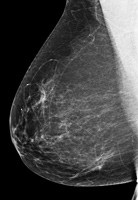
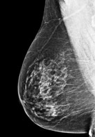
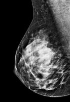
The mammograms below show a range of breast density. Some breasts are mostly fat (fatty breasts), some breasts are mostly breast tissue (dense breasts) and others are somewhere in between.
 |
 |
 |
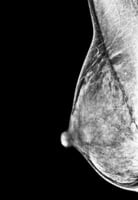 |
| Fatty breast | Some breast density | Dense breast | Very dense breast |
Images used with permission of the American College of Radiology (www.acr.org). |
|||
Breast density after menopause
Breast density may decrease after menopause in both women who go through natural menopause and younger women who are in menopause after surgery to remove the ovaries (oophorectomy).
For women who use menopausal hormone therapy (MHT), breast density may not decrease until they stop using MHT. MHT is also called postmenopausal hormone therapy and hormone replacement therapy (HRT).
Breast density and breast cancer risk
Women with dense breasts (as seen on a mammogram) have a somewhat higher risk of breast cancer than women with fatty breasts [19-20].
However, breast density does not appear to be related to a woman’s risk of dying from breast cancer [21-23].
Learn more about breast density and breast cancer risk.
Screening tests under study for women with dense breasts
Dense breast tissue can make abnormal findings hard to see on a mammogram. Screening tools are under study for women with dense breasts.
There are no special recommendations or breast cancer screening guidelines for women with dense breasts. However, your health care provider may suggest other types of breast imaging in addition to regular mammograms.
Breast ultrasound and breast MRI (each in combination with mammography) are under study for screening in women with dense breasts.
Breast density legislation
In 2019, the U.S. Congress passed national breast density legislation. The Food and Drug Administration (FDA) now requires all mammography centers to notify all patients about the density of their breasts after their mammograms. Centers must also notify health care providers about their patients’ breast density.
Although this may seem helpful, there are no special screening guidelines for women with dense breasts. If you have dense breasts, talk with your health care provider about the risks and benefits of getting other breast imaging in addition to regular mammograms.
If you have any concerns about your breast density or your risk of breast cancer, talk with your health care provider.
Read our blog, Understanding What it Means if You’re Told You Have Dense Breasts.
Findings on a mammogram
Benign (not cancer) breast conditions
Some common benign breast conditions, such as cysts and fibroadenomas, may show up on mammograms as round or oval patches with distinct borders. These changes are not related to cancer, and these findings are not abnormal.
Learn more about benign breast conditions.
Calcifications and microcalcifications
Calcifications are bits of calcium that can show up on mammograms as small, bright white spots.
Most calcifications are benign (not cancer). However, certain patterns of calcifications are suspicious and need more testing. Tight clusters or lines of microcalcifications (tiny calcifications) can be a sign of breast cancer.
Calcifications are common, especially after age 50 [24]. They may be related to past injury to the breast or mastitis (an infection in the breast) [24].
In women treated for breast cancer in the past, calcifications may be related to past breast surgery or radiation therapy [24].
Calcifications are not related to the amount of calcium in your diet [24].
Architectural distortion
Architectural distortion describes a distorted shape or pattern of breast tissue on a mammogram. However, no mass is seen. If you have architectural distortion, your radiologist may compare your mammogram to a past mammogram to see if the finding is new.
Although the breast tissue may be normal, architectural distortion usually needs follow-up testing because it may be a sign of a benign (not cancer) breast condition or breast cancer.
Ductal carcinoma in situ (DCIS)
Ductal carcinoma in situ (DCIS) is a non-invasive breast cancer. It’s stage 0 breast cancer.
On a mammogram, DCIS usually looks like a cluster of microcalcifications. It can be hard to know from a mammogram whether the cluster is DCIS or invasive breast cancer. A cluster of microcalcifications can also be a benign (not cancer) finding on a mammogram.
Learn about follow-up after an abnormal mammogram.
Learn more about DCIS.
Invasive breast cancer
Invasive breast cancer can look like a white patch or mass on a mammogram. Invasive breast cancers include invasive ductal cancer and invasive lobular cancer.
With invasive ductal cancer, the tumor cells that began in the milk duct don’t stay within the clear borders of the duct. Instead, they invade the nearby breast tissue. The outer edges of these cells look fuzzy or spiky (called spiculated).
With invasive lobular cancer, the tumor cells begin in the lobules (the milk-producing sacs) of the breast. These breast cancers can be harder to find on a mammogram.
Learn about follow-up after an abnormal mammogram.
The following is an interactive model of what a mammogram may show.
Mammography results
When to expect mammography results
Some centers give you the results of your mammogram at the time of your screening. With others, it may take up to 2 weeks to get your results.
If you don’t get your results within 2 weeks, contact your health care provider or the mammography center.
Don’t assume the results were normal because you didn’t get a report. Follow up to get your results.
Breast Imaging Reporting and Data System
Most mammography centers report the results of mammograms using the Breast Imaging Reporting and Data System (BI-RADS®).
BI-RADS® was developed by the American College of Radiology to provide a standard way to describe mammography findings (with categories numbered 0 to 6).
Assessment is complete |
|
BI-RADS® category |
What this means |
1: Negative |
No evidence of cancer on the mammogram. Nothing suspicious or worrisome was seen on the mammogram. |
2: Benign finding(s) |
No evidence of cancer on the mammogram. This is also a negative result, but to be complete it’s noted there are findings that appear benign (not suspicious), such as a cyst. |
3: Probably benign finding – initial short-interval follow-up suggested |
More than 98% of these will NOT be cancer, but to be sure, follow-up exams are needed (usually at 6 months and then afterwards as needed). |
4: Suspicious abnormality – biopsy should be considered |
The findings don’t definitely look like cancer but could be cancer. Some, but not all, radiologists may divide this category further into 4A, 4B and 4C based on how suspicious the findings are for cancer (4A looks the least like cancer). |
5: Highly suggestive of malignancy – appropriate action should be taken |
The finding has a high chance (95% or more) of being cancer. |
6: Known biopsy-proven malignancy – appropriate action should be taken |
This category is only for findings already confirmed as cancer with a biopsy. |
Assessment is incomplete |
|
BI-RADS® category |
What this means |
0: Need additional imaging evaluation and/or prior mammograms |
More information is needed to know whether a finding is abnormal. |
Adapted from American College of Radiology’s BI-RADS® – Mammography, Fifth Edition and American Cancer Society materials [25-26] with permission of the American College of Radiology. |
|
Updated 03/17/25
This content is regularly reviewed by an expert panel including researchers, practicing clinicians and patient advocates.