Core Needle Biopsy Procedure and Recovery
Core needle biopsy uses a hollow needle to remove samples of tissue from the breast. It’s the standard and preferred way to diagnose breast cancer. It may also rule out breast cancer.
A pathologist studies the tissue samples under a microscope to see if they contain cancer. If they do, more tests will be done to help plan your treatment.
Core needle biopsy can be used to check a:
- Lump that can be felt (palpable mass)
- Suspicious area that can only be seen on a mammogram or other imaging test (nonpalpable abnormal finding)
Learn about factors that affect your prognosis (chances of survival) and treatment.
Learn more about types of breast biopsies.
Core needle biopsy for a palpable mass
If you have a palpable mass (a lump that can be felt), a core needle biopsy may be done in your doctor’s office, a hospital or an imaging center.
Before the procedure begins, your doctor will use a small amount of local anesthetic to numb the skin and breast tissue around the suspicious area.
Your doctor will then insert the needle and remove a small amount of tissue. They may use images from a breast ultrasound to guide the needle to the suspicious area.
During a core needle biopsy, a clip may be placed inside the breast (you can’t feel it) to mark the location of the lump. The clip makes the lump easier to find if surgery is needed.
If the biopsy shows cancer, the clip will be removed during your breast surgery. If surgery is not needed (the biopsy shows no cancer), it’s safe to leave the clip in the breast.
Core needle biopsy for a nonpalpable abnormal finding
A non-palpable abnormal finding is a suspicious area that can’t be felt but can be seen on a mammogram or other imaging test. For example, it may be a mass or microcalcifications.
For non-palpable findings, a core needle biopsy is more involved than with lumps that can be felt. It’s done in a hospital or imaging center.
Your radiologist will use images from a breast ultrasound, breast MRI or a special type of mammography (called stereotactic mammography) to guide the needle to the suspicious area.
When the suspicious area can be seen on a breast ultrasound, breast ultrasound-guided biopsy is usually the preferred method [5].
During a core needle biopsy, a clip may be placed inside the breast (you can’t feel it) to mark the location of the suspicious area. The clip makes the suspicious area easier to find if surgery is needed.
If the biopsy shows cancer, the clip will be removed during your breast surgery. If surgery is not needed (the biopsy shows no cancer), it’s safe to leave the clip in the breast.
Core needle biopsy with breast ultrasound
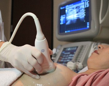
During a core needle biopsy with breast ultrasound, you lie on your back.
Before the procedure, your radiologist will use a local anesthetic to numb the area.
Your radiologist will hold the ultrasound device against your breast to see the area. The ultrasound images help your radiologist guide the biopsy probe to the suspicious area.

The ultrasound images help your radiologist guide the biopsy device to the suspicious area.
Your radiologist then removes a sample of tissue with the needle in the biopsy device. In some cases, this is done with a vacuum-assisted biopsy device. The needle is inserted and removed quickly.
You may feel a pushing and pulling sensation on your breast, which can cause discomfort.
The following is a 3D interactive model showing a core needle biopsy with breast ultrasound. Click the arrows to move through the model to learn more about this procedure.
Core needle biopsy with breast MRI
During a core needle biopsy with breast MRI, you lie on your stomach on a special table with a hole where your breast fits through.
Before the procedure, you will be given a contrast agent by vein (through an IV). Your radiologist will use a local anesthetic to numb the breast area.
Your breast will be compressed like it is for a mammogram, and several MRI images will be taken. These images help your radiologist guide the biopsy device to the suspicious area.
Your radiologist then removes a sample of tissue with the needle in the biopsy device. Usually, this is done with a vacuum-assisted biopsy device. The needle is inserted and removed quickly.
Your radiologist then removes a sample of tissue with the needle in a vacuum-assisted mechanism. The needle is inserted and removed quickly.
You may feel a pushing and pulling sensation on your breast, which can cause discomfort.
Core needle biopsy with stereotactic mammography
During a core needle biopsy with stereotactic mammography, you sit or lie on your stomach, depending on the imaging machine. With many imaging machines, you sit up and your breast is positioned much like it is for a mammogram. With other imaging machines, you lie down and your breast fits through a hole in the table. The biopsy is done from below the table.
Before the procedure, your radiologist will use a local anesthetic to numb the area.
Your breast will be compressed like it is for a mammogram, and several images are taken. Some imaging machines use digital breast tomosynthesis. The images help your radiologist guide the biopsy device to the suspicious area in the breast.
Your radiologist then removes a sample of tissue with the needle in the biopsy device. Usually, this is done with a vacuum-assisted biopsy device. The needle is inserted and removed quickly.
You may feel a pushing and pulling sensation against your breast, which can cause discomfort.
What to expect
After a core needle biopsy, you may feel sore in the biopsy area. Placing an ice pack on the area, resting, or taking a mild pain reliever such as ibuprofen (Advil or Motrin), naproxen (Aleve or Naprosyn) or acetaminophen (Tylenol) may help.
You may also feel a buildup of emotions. You may be anxious leading up to the procedure. You might feel discomfort or distress during the procedure. And you may be worried about the results.
You may want to take the day off from work and other commitments to recover both physically and emotionally. You may also want to have a family member or friend go with you to the procedure and be available afterwards for support.
Find more ways to cope while you wait for your biopsy results.
Advantages of core needle biopsy
Core needle biopsy is quick and doesn’t involve surgery. There’s only a small chance of infection or bruising.
If breast cancer is found, the tissue removed during a core needle biopsy gives important information including:
This information helps guide your treatment.
If the tissue sample is benign (not cancer), surgery may be avoided. In some cases, however, even if the tissue sample is benign, a surgical biopsy may be needed to confirm the diagnosis.
Drawbacks of core needle biopsy
One drawback of core needle biopsy is the needle can miss the tumor and take a sample of nearby normal tissue instead. This is most likely to occur when the biopsy is done without the help of breast ultrasound, breast MRI or stereotactic mammography.
If a tumor is missed, the biopsy will show cancer doesn’t exist when in fact, it does. This is called a false negative result and delays diagnosis.
- For abnormal findings that can’t be felt (can only be seen on a mammogram or other imaging test), false negative results occur in up to 4% of image-guided core needle biopsies [5-7].
- For masses that can be felt, false negative results occur less often than with abnormal findings that can’t be felt (can only be seen on a mammogram or other imaging test) [8].
Another drawback of core needle biopsy is that it may not give full information about the tumor. For example, it can’t tell the size of a tumor.
Taking multiple tissue samples may help limit this problem. However, in some cases, a surgical biopsy is needed to get complete information about the tumor.
Updated 06/05/23

